


Decoding the Diagnosis: The Power of Prostate MRI Reporting Templates for Accurate Prostate Cancer Detection
Prostate cancer is a serious health concern, and early detection is crucial for effective treatment. Modern medicine has advanced significantly, and one of the most important tools in diagnosing prostate cancer is the prostate MRI. However, the effectiveness of an MRI relies heavily on the quality of its interpretation. Enter: Prostate MRI reporting templates. These standardized frameworks guide radiologists and urologists, ensuring consistency, accuracy, and ultimately, improved patient outcomes. This article dives deep into the world of prostate MRI reporting templates, exploring their structure, benefits, and impact on the diagnostic process.
Understanding the Role of Prostate MRI in Prostate Cancer Diagnosis
Before exploring the templates themselves, it’s important to understand the significance of prostate MRI. This non-invasive imaging technique provides detailed images of the prostate gland, allowing doctors to:
- Visualize the prostate’s internal structure: Identifying any abnormalities like tumors or inflammation.
- Assess the size and location of suspicious lesions: Pinpointing the exact areas of concern.
- Determine the stage of the cancer: Helping to guide treatment decisions.
- Guide biopsies: Targeting the most suspicious areas for tissue sampling.
- Monitor treatment response: Assessing the effectiveness of therapies like radiation or chemotherapy.
The Evolution and Purpose of Prostate MRI Reporting Templates
The development of standardized reporting templates was a game-changer in prostate cancer diagnosis. Before these templates, reports were often inconsistent, making it difficult to compare results and accurately assess the risk of cancer. These templates provide a structured approach to reading and reporting prostate MRI findings, leading to:
- Increased Accuracy: By providing a systematic checklist, templates minimize the risk of overlooking important details.
- Improved Consistency: Standardization ensures that all radiologists and urologists interpret the images using the same criteria.
- Facilitated Communication: A common language makes it easier for specialists to share information and collaborate on patient care.
- Enhanced Patient Management: Accurate and consistent reports lead to more informed treatment decisions and improved patient outcomes.
The PI-RADS System: The Gold Standard in Prostate MRI Reporting
The Prostate Imaging-Reporting and Data System (PI-RADS) is the most widely used and recognized reporting system for prostate MRI. It’s a collaborative effort between the American College of Radiology (ACR), the European Society of Urogenital Radiology (ESUR), and the AdMeTech Foundation. PI-RADS uses a scoring system to assess the likelihood of clinically significant prostate cancer.
Key Features of the PI-RADS System:
- Standardized Protocol: Defines the specific MRI sequences required for a complete prostate MRI exam.
- Structured Reporting: Provides a standardized format for reporting findings, including lesion location, size, and characteristics.
- PI-RADS Score: Assigns a score from 1 to 5 based on the likelihood of clinically significant cancer:
- PI-RADS 1: Very low risk of clinically significant cancer.
- PI-RADS 2: Low risk of clinically significant cancer.
- PI-RADS 3: Equivocal – the presence of clinically significant cancer is uncertain.
- PI-RADS 4: High risk of clinically significant cancer.
- PI-RADS 5: Very high risk of clinically significant cancer.
- Recommendations: Guides recommendations for further evaluation, such as prostate biopsy.
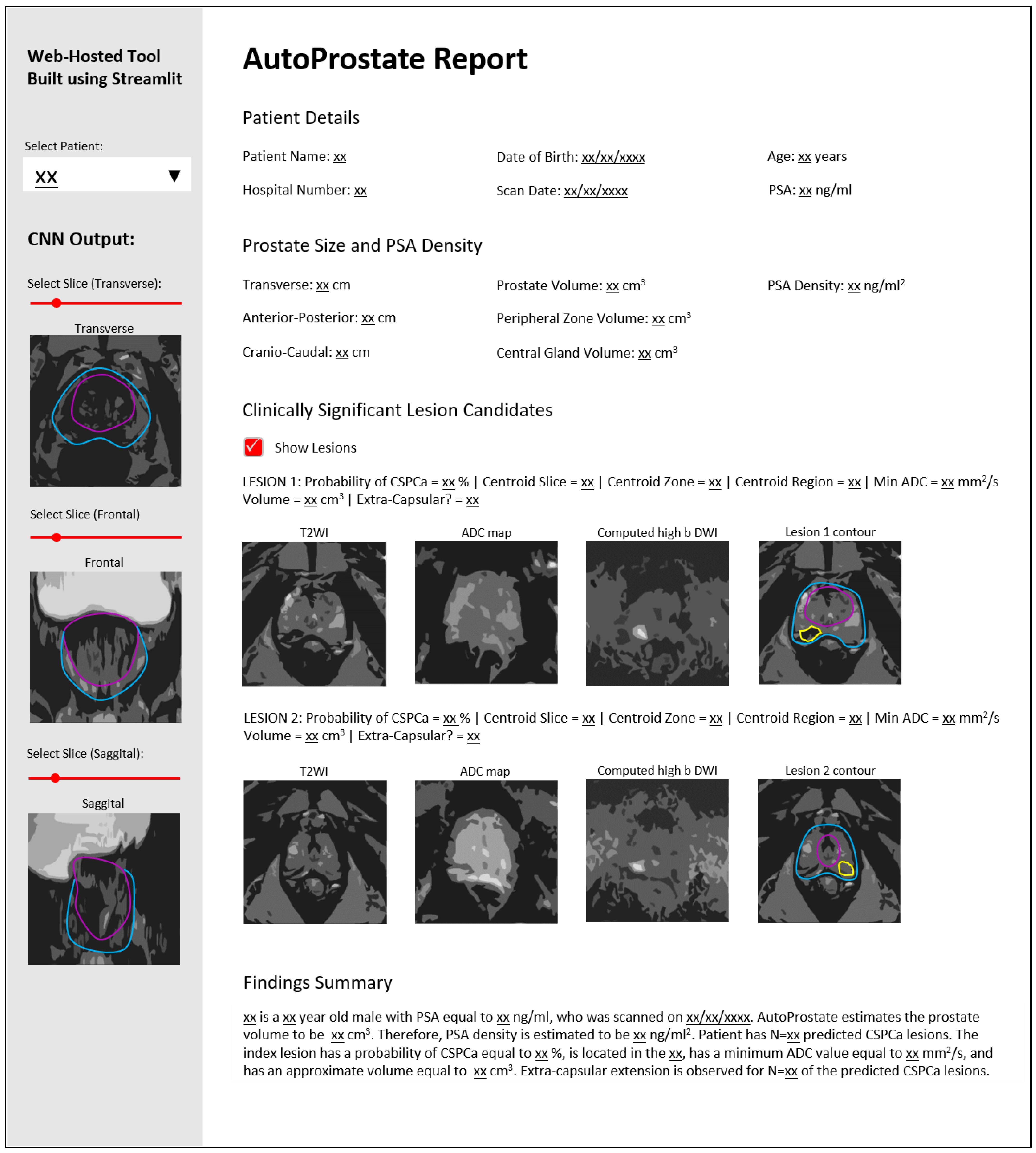
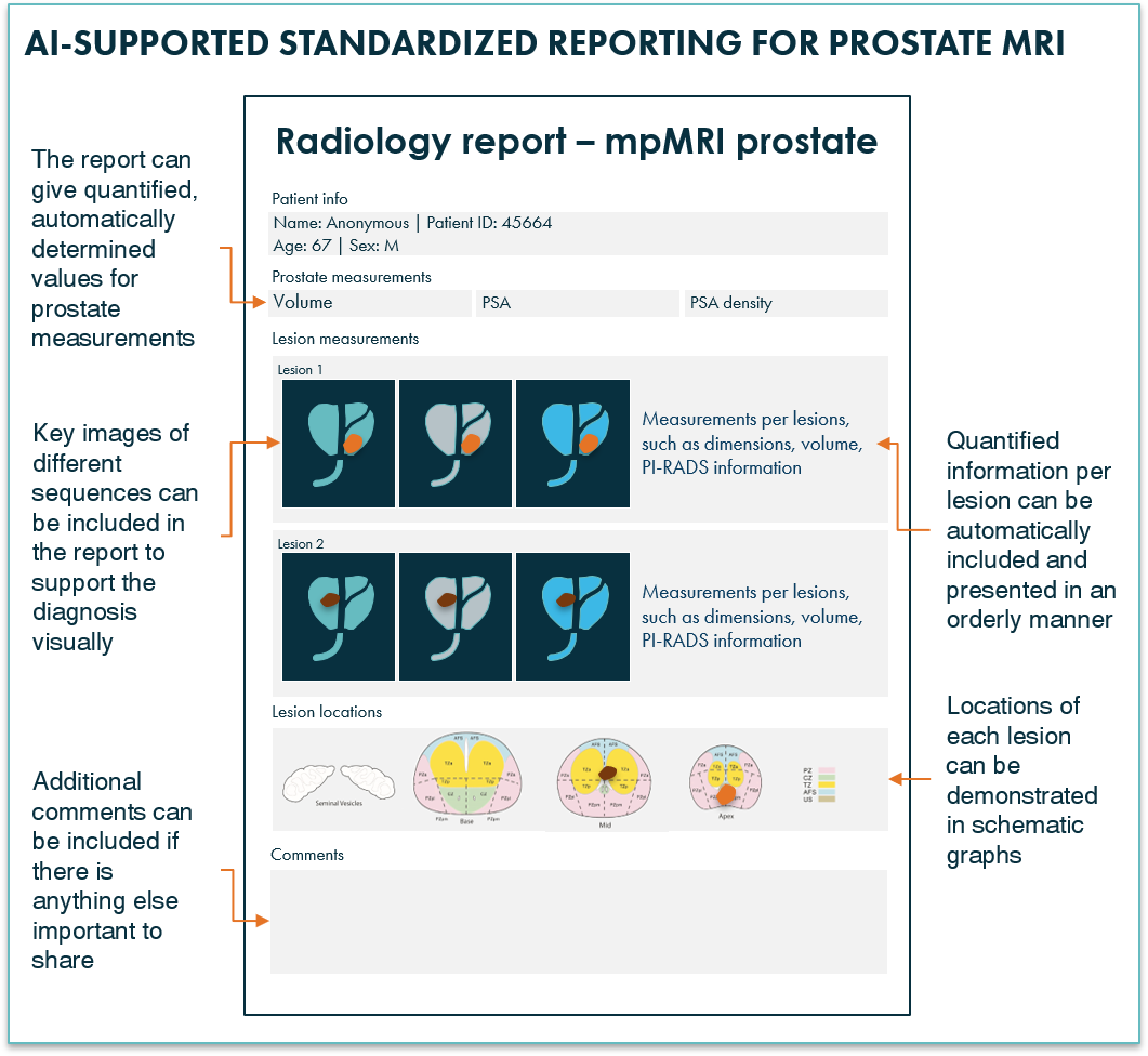
Components of a Typical Prostate MRI Reporting Template
While the PI-RADS system is the foundation, the specific format of a reporting template can vary. However, most templates include the following key components:
- Patient Demographics: Age, medical history, PSA levels, and prior biopsies.
- Technical Quality: Assessment of the image quality to ensure it meets the required standards.
- Anatomic Location: Description of the prostate zones (peripheral, central, and transition zones) and the location of any suspicious lesions within these zones.
- MRI Sequences: Evaluation of the findings on different MRI sequences, including T2-weighted imaging, diffusion-weighted imaging (DWI), and dynamic contrast-enhanced (DCE) imaging.
- PI-RADS Score: Assignment of a PI-RADS score based on the overall assessment.
- Overall Impression and Recommendations: A summary of the findings and recommendations for further management, such as biopsy or follow-up MRI.
Benefits of Using Reporting Templates for Doctors
The use of prostate MRI reporting templates offers numerous advantages for physicians involved in prostate cancer diagnosis and treatment:
- Improved Diagnostic Accuracy: Structured approach reduces the likelihood of errors.
- Enhanced Communication: Clear and consistent reports facilitate communication among specialists.
- Standardized Assessment: Ensures consistent interpretation regardless of the radiologist.
- Facilitates Research and Audit: Allows for the tracking of outcomes and continuous improvement.
- Improved Patient Outcomes: Leads to earlier and more accurate diagnoses, ultimately impacting patient survival and quality of life.
Conclusion: The Future of Prostate Cancer Diagnosis
Prostate MRI reporting templates, particularly the PI-RADS system, have revolutionized prostate cancer diagnosis. By providing a standardized framework for interpreting MRI images, these templates ensure accuracy, consistency, and improved communication among healthcare professionals. As technology continues to advance, these templates will likely evolve to incorporate new imaging techniques and refine the diagnostic process further. Ultimately, the use of prostate MRI reporting templates empowers physicians to make more informed decisions, leading to earlier detection, more effective treatment, and better outcomes for patients battling prostate cancer.
Frequently Asked Questions (FAQs)
What is the purpose of a PI-RADS score?
The PI-RADS score is a standardized rating system that helps radiologists and urologists assess the likelihood that a suspicious area on a prostate MRI represents clinically significant prostate cancer. The score ranges from 1 to 5, with higher scores indicating a greater likelihood of cancer.
What happens if my prostate MRI reveals a PI-RADS 4 or 5 score?
A PI-RADS 4 or 5 score suggests a high probability of clinically significant cancer. Your doctor will likely recommend a prostate biopsy to confirm the diagnosis and assess the aggressiveness of the cancer.
Are prostate MRI reporting templates used worldwide?
The PI-RADS system is widely adopted internationally. However, specific implementation and the use of other, more specialized reporting systems may vary slightly based on local guidelines and resources.
Can I request a copy of my prostate MRI report?
Yes, you have the right to request a copy of your medical records, including your prostate MRI report, from your healthcare provider.
